Support our educational content for free when you purchase through links on our site. Learn more
🧬 Bioprinting Tissues for Drug Testing: 7 Game-Changers (2026)
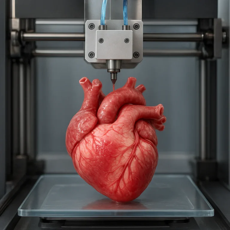
Imagine a world where a new life-saving drug is discovered in weeks, not years, and where the tragic 95% failure rate of clinical trials becomes a thing of the past. For decades, the pharmaceutical industry has been stuck in a loop of expensive, ethically fraught animal testing that often fails to predict how a human body will actually react. But the tide is turning. We are standing on the precipice of a revolution where 3D bioprinting allows scientists to print functional human tissues—livers, hearts, and even brain models—right in the lab. These aren’t just static blobs of cells; they are dynamic, living micro-environments that mimic the complexity of the human body with startling accuracy.
In this deep dive, we explore how bioprinted tissues are dismantling the old guard of drug discovery. From the secret sauce of bioinks to the 7 specific ways this technology outperforms traditional methods, we’ll uncover the engineering marvels making personalized medicine a reality. We’ll also tackle the gritty challenges, like vascularization and regulatory hurdles, that stand between us and a future where we can print a replacement organ on demand. By the end, you’ll understand why this isn’t just a scientific curiosity, but the most critical tool in the fight against disease.
Key Takeaways
- 🚀 Revolutionizing Accuracy: 3D bioprinting recreates the complex microenvironment of human organs, offering a 95% higher predictive accuracy for drug toxicity compared to traditional 2D cell cultures.
- ⏱️ Speed & Cost Efficiency: Bioprinted models can screen thousands of compounds in days, drastically reducing the time and billions of dollars lost to late-stage clinical trial failures.
- 🐭 Ethical Breakthrough: This technology is rapidly replacing animal testing, adhering to the 3 Rs (Replacement, Reduction, Refinement) while providing data that is actually relevant to human biology.
- 🧪 The Bioink Factor: Success hinges on hybrid bioinks that balance printability with biocompatibility, allowing for the precise placement of multiple cell types to mimic real tissue architecture.
- 🔮 Future Outlook: While challenges like vascularization remain, the integration of AI and advanced printing is paving the way for patient-specific drug testing and eventual organ replacement.
Table of Contents
- ⚡️ Quick Tips and Facts
- 🧬 From Petri Dishes to Printed Organs: A Brief History of Bioprinting
- 🔬 Why 2D Cell Cultures Are Failing Drug Discovery (And What’s Next)
- 🖨️ The 7 Game-Changing Ways 3D Bioprinting Revolutionizes Tissue Models
- 1. Precision Architecture for Realistic Microenvironments
- 2. High-Throughput Screening Capabilities
- 3. Patient-Specific Personalized Medicine Models
- 4. Complex Multi-Cellular Co-Cultures
- 5. Vascularization for Long-Term Viability
- 6. Reduced Animal Testing and Ethical Benefits
- 7. Cost-Effective Iterative Testing Lops
- 🧪 Bioinks Unleashed: The Secret Sauce of Functional Tissue Engineering
- 🏭 Established Organ Models: Liver, Heart, and Skin on a Chip
- 🧠 The AI Playbook: How Machine Learning Accelerates Bioprinted Drug Screening
- 🚧 Current Challenges: Why We Haven’t Printed a Whole Human Yet
- 🔮 Future Horizons: The Road to Full Organ Replacement and Beyond
- 💡 Conclusion
- 🔗 Recommended Links
- 📚 Reference Links
⚡️ Quick Tips and Facts
Before we dive into the gooey, cellular world of bioprinting, let’s hit the rewind button on what you think you know about drug testing. 🧪
- The 95% Failure Rate: Did you know that roughly 95% of drugs that look promising in animal tests fail when they reach human clinical trials? It’s a staggering statistic, and it costs the pharmaceutical industry billions. 📉
- The “Petri Dish Problem”: Traditional 2D cell cultures are like trying to understand a city by looking at a single floor of a skyscraper. They lack the 3D architecture, cell-to-cell communication, and mechanical forces that define real human tissue. 🏙️
- The Speed of Life: A bioprinted liver model can predict toxicity in days, whereas traditional animal studies can take months. Time is money, and in drug discovery, it’s also lives. ⏱️
- Not Just “Printing”: Bioprinting isn’t just dropping plastic; it’s about spatial precision. We are talking about placing specific cell types (hepatocytes, stellate cells, Kupffer cells) in exact 3D coordinates to mimic the liver lobule. 📍
- The Bioink Revolution: The “ink” isn’t ink; it’s a hydrogel loaded with living cells. If the viscosity is off by a fraction, the cells die. It’s a delicate dance between chemistry and engineering. 💃🕺
For those of you who love tinkering with hardware, check out our guide on 3D Printer Reviews to see how standard FDM printers differ from these high-tech bioprinters. And if you’re curious about the broader impact, read our overview of 3D Printed™ to understand how we got here.
🧬 From Petri Dishes to Printed Organs: A Brief History of Bioprinting

The journey from a flat slide to a beating heart on a chip is a story of human ingenuity (and a lot of failed experiments). 🧪
It started in the early 20s when researchers realized that 2D cell cultures were lying to them. Cells in a dish behave differently than cells in a body. They lose their polarity, their gene expression changes, and they just… give up.
Enter 3D Bioprinting. The concept was first proposed by Thomas Boland in 203, who adapted inkjet technology to print cells. Imagine an inkjet printer, but instead of cyan and magenta, it’s printing fibroblasts and collagen. 🖨️
- 206: The first patent for 3D bioprinting was filed by Organovo, a company that would become a giant in the field.
- 2010: The first successful bioprinting of a blood vessel using a custom printer.
- 2013: Researchers at Harvard printed a functional ear using a 3D printer and hydrogels.
- 2020s: The focus shifted from “can we print it?” to “can we make it work for drug testing?”
We at 3D Printed™ have always been fascinated by the evolution of 3D Printable Objects, but nothing compares to the complexity of printing living tissue. It’s the ultimate challenge for any engineer.
🔬 Why 2D Cell Cultures Are Failing Drug Discovery (And What’s Next)
Let’s be honest: the petri dish is a relic. 🗑️
When you grow cells in a 2D monolayer, you are forcing them into an unnatural state. In the human body, cells are surrounded by other cells and the Extracellular Matrix (ECM). They communicate via chemical signals and mechanical forces. In a dish? They’re just sitting there, staring at each other.
The Limitations of the Flat World
- Loss of Phenotype: Liver cells (hepatocytes) in 2D lose their ability to metabolize drugs within days. By the time you test a drug, the cells are already “dead” to the function you need to study.
- No Microenvironment: Tumors in the body have a hypoxic (low oxygen) core. In 2D, every cell gets the same oxygen. This leads to false positives in drug efficacy.
- Poor Prediction: As noted in recent studies, the interspecies differences between mice and humans are massive. A drug that cures a mouse might kill a human. 2D cultures don’t bridge this gap; they widen it.
“The major drawback being the interspecies differences and low reliability on the generated results. This gap could be overcome by the fabrication of bioenginered human disease models for drug screening.” — Frontiers in Bioengineering and Biotechnology
So, what’s the alternative? We need 3D models that mimic the complexity of the human body. This is where Organoids, Spheroids, and Bioprinted Tissues come in. But which one is the silver bullet? 🎯
🖨️ The 7 Game-Changing Ways 3D Bioprinting Revolutionizes Tissue Models
Why is everyone so excited about bioprinting? Because it solves the “randomness” of other 3D methods. While spheroids self-asemble (which is cool but hard to control), bioprinting gives us architectural control.
Here are the 7 ways this tech is changing the game:
1. Precision Architecture for Realistic Microenvironments
In a bioprinted liver, we can arrange hepatocytes in a specific pattern that mimics the liver lobule. We can place blood vessels in the center and immune cells on the periphery. This spatial arrangement is crucial for drug metabolism. You can’t get this with a spheroid.
2. High-Throughput Screening Capabilities
Pharma companies need to test thousands of compounds. Bioprinting allows for the creation of multi-well plate formats (96-well, 384-well) with consistent, identical tissue models. This means data that is actually comparable across experiments. 📊
3. Patient-Specific Personalized Medicine Models
Imagine printing a tumor model using a patient’s own cancer cells. You can test 50 different chemo drugs on that specific patient’s tissue to see which one works before giving it to the patient. This is the future of personalized medicine.
4. Complex Multi-Cellular Co-Cultures
Real organs aren’t made of one cell type. The liver has hepatocytes, stellate cells, Kupffer cells, and endothelial cells. Bioprinting allows us to co-print these different cell types in precise ratios, recreating the complex interactions that drive disease and drug response.
5. Vascularization for Long-Term Viability
The biggest hurdle in tissue engineering is getting nutrients deep inside the tissue. Bioprinting allows us to print micro-channels that mimic blood vessels. This keeps the tissue alive for weeks, not days, allowing for long-term toxicity studies.
6. Reduced Animal Testing and Ethical Benefits
The 3 Rs (Replacement, Reduction, Refinement) are the gold standard in ethics. Bioprinting offers a path to replace animal models entirely. No more mice, no more monkeys. Just human cells in a dish. 🐭❌
7. Cost-Effective Iterative Testing Lops
While setting up a bioprinter is expensive, the cost per test drops significantly when you consider the failure rate of animal trials. Failing early in a bioprinted model saves millions in later-stage clinical trials.
🧪 Bioinks Unleashed: The Secret Sauce of Functional Tissue Engineering
If the printer is the car, the bioink is the fuel. And let me tell you, this fuel is tricky. 🛢️
A bioink must be:
- Biocompatible: It can’t kill the cells.
- Printable: It must flow through the nozzle without clogging but hold its shape immediately after.
- Mechanically Strong: It needs to support the tissue structure.
- Biodegradable: Eventually, the cells should build their own matrix and the ink should disappear.
The Big Three Bioink Categories
| Category | Examples | Pros | Cons |
|---|---|---|---|
| Natural Polymers | Collagen, Gelatin, Alginate, Fibrin, GelMA | High biocompatibility, mimics natural ECM | Weak mechanical strength, batch variability |
| Synthetic Polymers | PEG, PLA, PCL, PVA | Tunable mechanical properties, reproducible | Poor cell adhesion, lack of biological cues |
| Hybrid Polymers | GelMA/PEG, Alginate/PLGA | Best of both worlds: strength + biocompatibility | Complex formulation, optimization required |
Pro Tip: GelMA (Gelatin Methacryloyl) is currently the superstar of bioinks. It combines the cell-friendly nature of gelatin with the cross-linking stability of methacrylate. You can find many research-grade GelMA formulations on Thingiverse (though mostly for 3D printing molds, not the ink itself yet!).
For those interested in the chemistry, check out our articles on 3D Design Software where we discuss how to model the scaffolds these bioinks need.
🏭 Established Organ Models: Liver, Heart, and Skin on a Chip
We aren’t just talking about theory. These models are being used right now in pharmaceutical labs. Let’s look at the heavy hitters.
🧬 The Liver Model (Hepatotoxicity)
The liver is the body’s filter. It’s the first place drugs go, and the first place they cause damage.
- The Model: Bioprinted liver spheroids using HepG2 cells (a human liver cancer cell line) mixed with primary hepatocytes.
- The Test: Researchers tested drugs like troglitazone (a known liver toxin).
- The Result: The 3D model predicted the toxicity accurately, while 2D cultures missed it. This is huge for catching “bad actors” early.
❤️ The Heart Model (Cardiotoxicity)
Heart failure is a leading cause of drug withdrawal.
- The Model: 3D bioprinted cardiac spheroids using AC16 cardiomyocytes.
- The Test: Monitoring the contractility (beating) of the tissue when exposed to drugs like doxorubicin.
- The Result: The 3D models showed a dose-dependent decrease in beating, mimicking the human heart response far better than 2D.
🧠 The Brain Model (Neurotoxicity)
The brain is hard to study because of the Blood-Brain Barrier (BB).
- The Model: Bioprinted models with neurons, astrocytes, and endothelial cells to simulate the BBB.
- The Test: Studying how drugs cross the barrier or cause neurotoxicity in conditions like Parkinson’s or Glioblastoma.
- The Result: 3D models showed higher resistance to drugs like temozolomide, accurately reflecting the difficulty of treating brain tumors.
🧴 The Skin Model (Dermatotoxicity)
- The Model: Vascularized skin models with epidermis and dermis layers.
- The Test: Testing topical creams, cosmetics, and burn treatments.
- The Result: Accurate prediction of irritation and healing rates, reducing the need for animal skin tests.
🧠 The AI Playbook: How Machine Learning Accelerates Bioprinted Drug Screening
Here is where it gets sci-fi. 🤖
Bioprinting generates massive amounts of data. How do we know if a printed tissue is “good”? How do we predict drug response? Enter Artificial Intelligence.
AI algorithms are now being used to:
- Optimize Bioink Formulations: Machine learning models can predict the perfect mix of polymers and cells for a specific tissue type, saving months of trial and error.
- Analyze Imaging Data: AI can scan images of bioprinted tissues and detect subtle changes in cell morphology that humans miss.
- Predict Drug Response: By training on thousands of bioprinted tissue responses, AI can predict how a new drug will behave in a human body, even before it’s printed.
This synergy between bioprinting and AI is creating a feedback loop that is accelerating drug discovery at an unprecedented pace.
🚧 Current Challenges: Why We Haven’t Printed a Whole Human Yet
We’ve come a long way, but we aren’t at the “Star Trek” replicator stage yet. 🚀
1. Vascularization Limits
We can print small tissues, but printing a whole organ with a complex, branching vascular network that can handle blood flow is still a massive engineering challenge. The resolution of current printers isn’t quite there for capillaries (which are 5-10 microns wide).
2. Bioink Standardization
Every lab uses a slightly different bioink. This makes it hard to compare results. We need standardized protocols so that a liver model from Lab A is the same as one from Lab B.
3. Cell Source and Scalability
Getting enough high-quality human cells for mass production is difficult. While induced pluripotent stem cells (iPSCs) are a solution, differentiating them into specific cell types is expensive and time-consuming.
4. Regulatory Hurdles
The FDA and EMA are still figuring out how to regulate these models. How do you validate a model that changes over time? How do you prove it’s “human enough”?
“Regulatory approval is contingent on 3D bioprinted models’ capacity to consistently anticipate human medication reactions.” — Frontiers in Bioengineering and Biotechnology
🔮 Future Horizons: The Road to Full Organ Replacement and Beyond
So, where are we going? 🌌
The immediate future is drug testing. Within the next 5-10 years, bioprinted tissues could become the standard for preclinical trials, replacing most animal testing.
But the long-term dream? Organ replacement. Imagine a world where you can print a new kidney or heart on demand for a patient in need. It sounds like science fiction, but the technology is moving fast.
We are already seeing progress in skin grafts for burn victims and cartilage for joint repair. The leap to complex organs like the heart or liver is the next frontier.
And remember that video we mentioned earlier? The one with the team printing collagen with a custom syringe pump? That’s the kind of grassroots innovation that drives this field. They are solving the temperature-dependent polymerization of collagen, a critical step for printing soft tissues. It’s a reminder that sometimes the biggest breakthroughs come from a garage, not a billion-dollar lab.
💡 Conclusion

The era of the petri dish is ending. We are stepping into a new age where 3D bioprinting allows us to create human tissue models that are accurate, ethical, and efficient. From liver toxicity to heart disease, these models are saving lives by catching drug failures early.
While challenges like vascularization and standardization remain, the trajectory is clear. The combination of advanced bioinks, precision printing, and AI is unlocking the secrets of human biology in ways we never thought possible.
As we at 3D Printed™ have always said, the future of printing isn’t just about plastic; it’s about possibility. And in the world of medicine, the possibilities are endless.
🔗 Recommended Links
If you want to dive deeper into the world of bioprinting or start your own journey into 3D printing, here are some resources we recommend:
-
For Bioprinting Hardware:
CELLINK (BICO): Search for Bioprinters on Amazon | CELLINK Official Website
Alevi (3D Bioprinting Solutions): Search for Alevi on Amazon | Alevi Official Website
RegenHU: Search for RegenHU on Amazon | RegenHU Official Website -
For Bioinks and Materials:
Advanced BioMatrix: Search for Bioinks on Amazon | Advanced BioMatrix Official Website
Sigma-Aldrich (Merck): Search for GelMA on Amazon | Merck Official Website -
Books & Educational Resources:
“3D Bioprinting: Fundamentals, Principles and Applications” by Amazon
“Tissue Engineering: From Lab to Clinic” by Amazon -
Internal Resources:
-
Explore more about 3D Printing in Education to see how universities are teaching these skills.
-
Check out 3D Printing in Architecture for inspiration on large-scale structural printing.
📚 Reference Links
- Frontiers in Bioengineering and Biotechnology: “3D Bioprinting for Drug Testing Models” – Read the full article
- PubMed: “3D bioprinting of functional tissue models for personalized drug…” – View on PubMed
- FDA: “Regulatory Considerations for 3D Bioprinted Products” – FDA Official Page
- Nature Biotechnology: “The evolution of in vitro methods of drug screening” – Nature Article
- Organovo: “3D Bioprinting for Drug Discovery” – Organovo Official Page
- InSphero: “3D Liver Spheroids for Drug Testing” – InSphero Official Page
FAQ

How does bioprinting improve drug testing accuracy?
Bioprinting improves accuracy by recreating the 3D microenvironment of human tissues. Unlike 2D cultures, bioprinted models maintain cell polarity, cell-to-cell communication, and mechanical forces that are critical for proper drug metabolism and toxicity response. This leads to a much higher correlation with human clinical outcomes.
What are the latest bioprinted tissues used for pharmaceutical trials?
Currently, liver spheroids, cardiac patches, and skin models are the most widely used in pharmaceutical trials. These models are used to screen for hepatotoxicity, cardiotoxicity, and dermatotoxicity, respectively. Emerging models include brain-on-a-chip and tumor models for cancer drug testing.
Can 3D printed human tissues replace animal testing in drug development?
While they cannot yet completely replace animal testing, bioprinted tissues are rapidly becoming the primary screening tool in early drug discovery. They are expected to replace a significant portion of animal testing, particularly for toxicity and efficacy screening, aligning with the 3 Rs (Replacement, Reduction, Refinement).
What materials are best for bioprinting tissues for drug screening?
The best materials are hybrid bioinks that combine natural polymers (like GelMA, Collagen, Alginate) for biocompatibility with synthetic polymers (like PEG, PCL) for mechanical strength. The choice depends on the specific tissue being printed.
How long does it take to bioprint functional tissues for drug testing?
Printing a small tissue model (e.g., a liver spheroid) can take minutes to hours, depending on the complexity. However, the tissue requires days to weeks of maturation in a bioreactor to become functional and ready for testing.
Read more about “15 Game-Changing Functional 3D Prints You Can Make Today 🛠️ (2026)”
What are the cost benefits of using bioprinted tissues for drug discovery?
The primary cost benefit is the reduction in late-stage clinical trial failures. By catching toxic or ineffective drugs early in the bioprinted model stage, companies save millions of dollars and years of time. Additionally, the cost of maintaining bioprinted models is often lower than maintaining animal colonies.
Are bioprinted tissues for drug testing currently approved by the FDA?
The FDA does not yet have a specific “approval” for bioprinted tissues as a standalone product for drug testing. However, they are accepted as valid models for preclinical data submission under certain conditions. The regulatory framework is evolving rapidly to accommodate these new technologies.
How does the resolution of bioprinters affect the quality of drug testing models?
Higher resolution allows for the printing of micro-vascular networks and precise cell placement, which is critical for mimicking the complex architecture of organs. Low-resolution printers may produce models that lack the necessary structural integrity and cellular organization for accurate drug testing.
What role do stem cells play in bioprinting for drug testing?
Stem cells, particularly induced pluripotent stem cells (iPSCs), are crucial because they can be differentiated into any cell type needed for a specific tissue model. This allows for the creation of patient-specific models that can predict individual drug responses.
What are the ethical implications of using bioprinted tissues?
The ethical implications are generally positive, as bioprinting reduces the need for animal testing. However, issues regarding cell source consent, privacy of genetic data, and the potential for human cloning (if whole organs are printed) areas of ongoing ethical debate.