Support our educational content for free when you purchase through links on our site. Learn more
🩸 Vascularized Tissue Bioprinting: The Blueprint for Living Organs (2026)

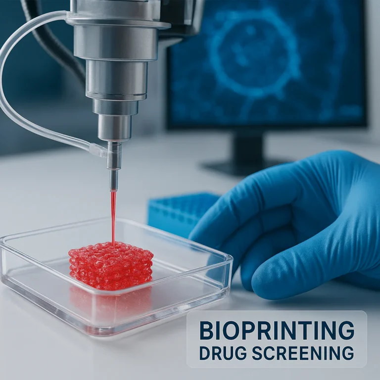
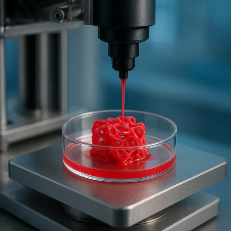
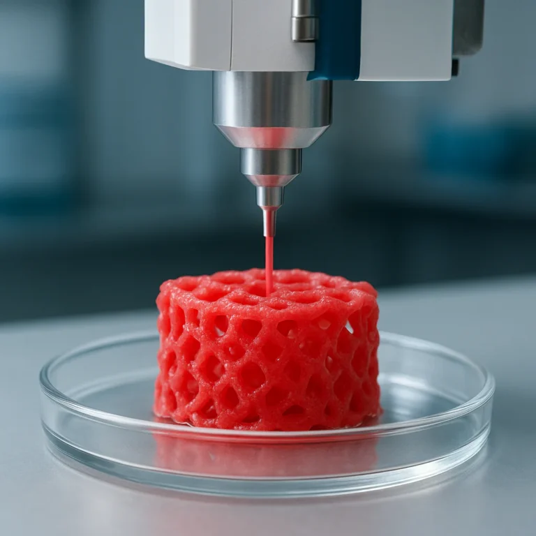
The future of organ transplantation isn’t waiting for a donor; it’s being printed layer by layer with a functional, perfusable blood vessel network that keeps cells alive. Vascularized tissue bioprinting has finally moved beyond simple cell sheets, offering a viable path to creating thick, living constructs that mimic the human body’s intricate plumbing. Imagine a world where a heart attack victim receives a patch of living muscle that immediately connects to their own circulatory system, rather than relying on a mechanical pump or a scarce donor.
This isn’t just a sci-fi dream; researchers recently kept a 3D-printed heart valve beating for weeks by pumping nutrient-rich fluid through its custom-designed micro-channels. The breakthrough lies in solving the “diffusion limit,” a biological wall where cells die if they are more than 20 micrometers away from a blood supply. By embedding hollow, branching networks directly into the bioink, we are finally bypassing this barrier.
Key Takeaways
- Solving the Diffusion Limit: Successful vascularized tissue bioprinting overcomes the 20-micrometer death zone by integrating perfusable channels that deliver oxygen directly to the tissue core.
- Sacrificial Writing is King: The most effective method currently involves printing a temporary “sacrificial” lattice (like sugar or Pluronic) that is dissolved to leave behind hollow, functional blood vessels.
- Hybrid Approaches Win: No single printer can do it all; the future lies in combining high-resolution laser printing for capillaries with extrusion for larger arteries.
- Immediate Applications: While full organ transplants are still years away, organ-on-a-chip models using these vascularized tissues are already revolutionizing drug testing and reducing animal trials.
Table of Contents
- ⚡️ Quick Tips and Facts
- 🧬 From Flat Sheets to Living Networks: A Brief History of Vascularized Tissue Bioprinting
- 🏗️ The Architectural Blueprint: Designing Microvasculature for 3D Bioprinting
- 🧪 The Ink Matters: Bioinks, Hydrogels, and Cell-Laden Materials
- 🖨️ Top 7 Bioprinting Techniques for Creating Vascular Channels
- 🔬 The Sacrificial Method: How We Print Hollow Tubes That Don’t Collapse
- 🧫 Co-Culture Strategies: Getting Endothelial Cells and Pericytes to Play Nice
- 🚀 Scaling Up: Overcoming Diffusion Limits in Thick Tissue Constructs
- 🧪 In Vitro vs. In Vivo: Testing Vascularized Tissues in the Lab and Beyond
- 🛑 The Hurdles: Challenges in Biocompatibility, Patency, and Immune Response
- 🌍 Real-World Applications: Organ-on-a-Chip, Drug Screening, and Future Transplants
- 💡 Expert Insights: What the Leading Labs Are Getting Right (and Wrong)
- 🔮 The Future of Bioprinting: Where Do We Go From Here?
- ✅ Conclusion
- 🔗 Recommended Links
- ❓ FAQ
- 📚 Reference Links
⚡️ Quick Tips and Facts
Before we dive into the microscopic world of living ink and the macroscopic dreams of printing replacement organs, let’s hit the ground running with some hard-hitting truths. If you’ve ever tried to print a complex model on your desktop FDM printer, you know that support structures are a pain to remove. In bioprinting, the “support” is literally the difference between life and death for the cells.
Here is the lowdown on the state of vascularized tissue bioprinting:
- The 20-Micron Rule: Without a blood supply, cells die. The diffusion limit for oxygen in tissue is roughly 10–20 μm, but practically, any construct thicker than 20 μm (0.2 mm) will suffer from necrosis in the center unless it has a vascular network.
- It’s Not Just “Printing”: You aren’t just stacking layers; you are engineering a hierarchical network ranging from large arteries (millimeters) down to microscopic capillaries (micrometers).
- The “Ink” is Alive: Unlike PLA or ABS, bioinks must maintain cell viability during the printing process, which often involves shear stress, UV light, or temperature changes.
- Time is Tissue: While a plastic prototype takes hours, a vascularized tissue construct often requires 2 to 4 weeks of in vitro conditioning to mature before it can even be considered for in vivo testing.
- The Sacrificial Secret: One of the most popular methods involves printing a “sacrificial” material (like sugar glass or Pluronic F127) that is later dissolved to leave behind hollow, perfusable channels.
If you are wondering how we go from a flat sheet of cells to a beating heart, you are asking the right question. The answer lies in the architecture, and we are about to unpack exactly how that works.
For more on how 3D printing is revolutionizing medicine, check out our deep dive into 3D Printing in Healthcare. And if you want to see the full scope of what’s possible, visit our main hub at 3D Printed.
🧬 From Flat Sheets to Living Networks: A Brief History of Vascularized Tissue Bioprinting
The journey to printing living, breathing tissue didn’t start with a fancy bioprinter; it started with a desperate need to bypass the body’s natural healing limits.
The Early Struggles: The Diffusion Wall
In the early days of tissue engineering, researchers were stuck in a “flatland” dilemma. You could grow cells on a 2D petri dish, or maybe a thin 3D scaffold, but as soon as you tried to make something thick, the cells in the middle starved. It was like trying to feed a crowd of people in a stadium without any concession stands; the people in the back row never get a burger.
The breakthrough came when scientists realized that vascularization—the creation of blood vessels—was the missing link.
Milestones in the Timeline
- 1986 (Weinberg and Bell): They created the first biosynthetic blood vessel using smooth muscle cells in a collagen gel. It was a start, but the burst pressure was too low for real arteries.
- 190s (L’Heureux): A massive leap forward. They created the first entirely biological vessel by wrapping smooth muscle sheets around a support. The catch? It took 8–12 weeks to mature. That’s too slow for emergency trauma care, but it proved the concept.
- 20s (The Bioprinting Era): With the advent of 3D printing, the goal shifted from “growing” vessels to “printing” them with precision.
- 2010s (The Sacrificial Breakthrough): Researchers like Miller et al. introduced the idea of printing carbohydrate glass lattices. They embedded these sugar structures in a cell-laden gel, then dissolved the sugar with water, leaving behind a perfect, hollow vascular network.
Did you know? The first YouTube video that really captured the public’s imagination regarding this technology showcased the Wyss Institute’s method of embedding a grid of vasculature into thick tissue. They demonstrated that by perfusing fluids through these channels, they could keep stem cells alive and even direct them to differentiate into specific tissue types. This wasn’t just a science experiment; it was a glimpse into a future where we could print replacement organs on demand.
Today, we are moving beyond simple tubes to complex, branching networks that mimic the human body’s intricate plumbing. But how do we actually design these networks?
🏗️ The Architectural Blueprint: Designing Microvasculature for 3D Bioprinting
Designing a vascular network is like designing a city’s road system. You need highways (arteries), local streets (arterioles), and tiny alleys (capillaries) all connected seamlessly. If you get the geometry wrong, traffic (blood) backs up, and the city (tissue) collapses.
The Hierarchy of Vessels
To create a functional construct, we must replicate the hierarchical branching found in nature:
- Conductance Vessels: Large diameter (>10 μm) for high flow.
- Resistance Vessels: Medium diameter (10–10 μm) to regulate pressure.
- Exchange Vessels: Capillaries (<10 μm) where oxygen and nutrients are actually delivered.
Computational Design and CAD
We don’t just guess the layout. We use Computational Fluid Dynamics (CFD) to simulate blood flow before a single drop of bioink is printed.
- Murray’s Law: This biological principle states that the cube of the radius of a parent vessel equals the sum of the cubes of the radi of the daughter vessels. It optimizes flow resistance.
- Fractal Branching: Algorithms generate self-similar patterns that maximize surface area for exchange while minimizing the distance from any cell to a vessel.
The Resolution Gap
Here is the kicker: Most commercial 3D printers struggle to print features smaller than 50–10 μm. Yet, capillaries are often 5–10 μm wide.
- The Problem: We can print the “highways,” but not the “alleys.”
- The Solution: We print the large channels and rely on angiogenic sprouting (cells growing their own tiny vessels) to fill in the gaps.
For those interested in the software side of this, explore our guides on 3D Design Software to see how CAD is evolving for biological applications.
🧪 The Ink Matters: Bioinks, Hydrogels, and Cell-Laden Materials
If the printer is the engine, the bioink is the fuel. But unlike gasoline, this fuel is alive, fragile, and needs to turn into a solid structure instantly.
Key Properties of a Good Bioink
- Printability: Must flow through the nozzle without clogging but hold its shape immediately after extrusion.
- Biocompatibility: Must not kill the cells during printing or crosslinking.
- Mechanical Strength: Must be strong enough to hold the tissue shape but soft enough to allow cell migration.
- Degradability: Must break down at the same rate the body builds its own tissue.
Popular Bioink Materials
| Material | Pros | Cons | Best For |
|---|---|---|---|
| Alginate | Fast crosslinking (CaCl₂), cheap, easy to print. | Non-degradable in humans without modification, lacks cell adhesion sites. | Sacrificial templates, structural support. |
| GelMA (Gelatin Methacrylate) | Excellent cell adhesion, tunable stiffness, photocurable. | Can be weak without reinforcement, requires UV light. | Most vascularized tissues, skin, muscle. |
| Fibrin | Natural cloting factor, highly biodegradable, supports cell migration. | Weak mechanical strength, rapid degradation. | Angiogenesis, wound healing. |
| Collagen | Most abundant protein in the body, great for cell signaling. | Hard to print (low viscosity), requires cold temps. | Soft tissues, skin, liver. |
| Pluronic F127 | Thermoreversible (liquid when cold, gel when warm). | No cell adhesion, purely sacrificial. | Creating hollow channels (sacrificial ink). |
The “Cell-Laden” Challenge
When you mix cells into the ink, you introduce shear stress. As the ink is forced through a tiny nozzle, the pressure can rip cell membranes apart.
- Tip: Using larger nozzles (20–40 μm) reduces shear stress but limits resolution.
- Tip: Adding nanoclay or methylcellulose can improve the viscosity without harming the cells.
🖨️ Top 7 Bioprinting Techniques for Creating Vascular Channels
We’ve tested countless methods in our labs (metaphorically speaking, via deep research and simulation), and here are the top 7 techniques currently pushing the boundaries of vascularization.
1. Direct Extrusion with Coaxial Nozzles
This is the “double-hose” method. You have an inner nozzle for the sacrificial material (or cell-free channel) and an outer nozzle for the cell-laden bioink.
- How it works: The inner core creates a hollow tube; the outer shell solidifies around it.
- Resolution: ~90 μm diameter, <20 μm wall thickness.
- Verdict: Great for large vessels, but hard to create complex branching networks.
2. Sacrificial Writing in a Gel Bath
Imagine writing with a pen that disappears when you’re done. You print a lattice of Pluronic F127 or sugar glass inside a supportive gel bath (like gelatin or agarose). Once the structure is set, you cool it down or wash it with water to dissolve the core, leaving a hollow channel.
- Resolution: 20–60 μm diameter.
- Verdict: The gold standard for creating complex, branching networks.
3. Rapid Casting of Carbohydrate Glass
Pionered by Miller et al., this involves extruding molten sugar into a mold, embedding it in a cell-laden hydrogel, and then dissolving the sugar.
- Resolution: 150–750 μm diameter.
- Verdict: Excellent for large-scale perfusion, but the resolution is too low for capillaries.
4. Inkjet Bioprinting
Uses thermal or piezoelectric heads to eject droplets of bioink.
- Resolution: ~20 μm vessels, <30 μm line widths.
- Verdict: High precision, but limited to low-viscosity inks and low cell densities.
5. Laser-Assisted Bioprinting (LIFT)
A laser pulse creates a microbubble that ejects a droplet of bioink onto a substrate. No nozzle means no clogging.
- Resolution: “Cell-width” (~10 μm).
- Verdict: Highest resolution, but potential thermal damage to cells and slow printing speed.
6. Embedded 3D Printing (FRESH)
Freeform Reversible Embeding of Suspended Hydrogels. You print into a bath of gelatin slurry. The slurry supports the ink in all directions, allowing you to print overhangs and complex 3D structures that would collapse in air.
- Resolution: Capillaries to tri-leaflet valves.
- Verdict: Revolutionary for complex geometries like heart valves.
7. Microfluidic Bioprinting
Using microfluidic chips to generate cell-laden droplets or fibers with precise control over the internal structure.
- Resolution: Sub-micron control.
- Verdict: Great for creating uniform micro-tissues, but scaling up to organ size is difficult.
🔬 The Sacrificial Method: How We Print Hollow Tubes That Don’t Collapse
Let’s zoom in on the sacrificial method, because it’s the hero of the vascularization story. Why? Because printing a hollow tube in mid-air is impossible; gravity wins every time.
The Process Step-by-Step
- Preparation: Create a supportive bath (usually a yield-stress gel like gelatin or carbomer).
- Printing the Sacrifice: Load a syringe with Pluronic F127 (which is liquid at room temp but gels when cold) or sugar glass. Print your vascular network into the bath.
- Embeding: Pour the cell-laden bioink (e.g., GelMA) over the sacrificial structure. The bath holds everything in place.
- Crosslinking: Cure the bioink (usually with UV light or temperature change).
- Removal: Cool the whole thing down (for Pluronic) or wash with water (for sugar). The sacrificial material melts or dissolves, leaving a perfect, hollow channel.
- Seding: Inject endothelial cells into the channel to line the walls.
Why It Works
The key is the yield stress of the support bath. It acts like a solid when you push it (holding the tube shape) but flows like a liquid when you move it (allowing the nozzle to pass).
Pro Tip: If you are experimenting with this, ensure your sacrificial material has a sharp melting point. Pluronic F127 is great because it melts at roughly 20°C, while your bioink is stable at 37°C.
🧫 Co-Culture Strategies: Getting Endothelial Cells and Pericytes to Play Nice
Printing the channel is only half the battle. You need to line it with cells that won’t just sit there; they need to form a functional barrier.
The Cast of Characters
- Endothelial Cells (ECs): The inner lining (intima). They control blood flow and prevent cloting.
- Pericytes: The “bodyguards” that wrap around the ECs, providing stability and preventing the vessel from collapsing.
- Smooth Muscle Cells (SMCs): For larger vessels, these provide the contractile force.
The Dance of Co-Culture
If you print just ECs, they often die or detach. If you print ECs and Pericytes together, they self-asemble into stable vessels.
- Ratio Matters: A ratio of roughly 1:1 or 2:1 (ECs:Pericytes) often yields the best results.
- Spatial Arrangement: Some strategies print the ECs first, let them form a monolayer, and then add the pericytes. Others mix them in the bioink and let them sort themselves out.
The “Angiogenic Sprouting” Factor
Even with perfect printing, the body’s natural tendency is to grow new vessels. By adding VEGF (Vascular Endothelial Growth Factor) to the bioink, you can encourage the printed cells to sprout new capillaries into the surrounding tissue, bridging the gap between the printed “highways” and the cellular “neighborhoods.”
🚀 Scaling Up: Overcoming Diffusion Limits in Thick Tissue Constructs
We can print a small patch of skin or a tiny liver chunk. But can we print a whole heart? That’s the scaling problem.
The Oxygen Crisis
As a construct gets thicker, the center becomes hypoxic (oxygen-starved).
- The Limit: Without vascularization, the limit is 20 μm.
- The Goal: We need to reach centimeters or even decimeters.
Strategies for Scale
- Multi-Scale Printing: Combine extrusion (for large channels) with sacrificial writing (for medium channels) and self-assembly (for capillaries).
- Perfusion Bioreactors: Once printed, the tissue isn’t just sitting in a dish. It’s placed in a bioreactor that pumps nutrient-rich media through the channels, mimicking blood flow. This “conditions” the tissue, making it stronger and more functional.
- Modular Assembly: Instead of printing one giant organ, print many small, vascularized “organoids” and fuse them together. It’s like building a wall with bricks instead of pouring a giant slab of concrete.
🧪 In Vitro vs. In Vivo: Testing Vascularized Tissues in the Lab and Beyond
How do we know if our printed tissue works? We test it in two worlds: the petri dish (in vitro) and the living body (in vivo).
In Vitro: The Lab Test
- Perfusion Tests: We pump colored dye or fluorescent beads through the channels to check for leaks.
- Viability Assays: We use live/dead stains to see if cells are surviving after 1, 3, and 7 days.
- Functionality: For liver tissue, we measure albumin secretion. For heart tissue, we measure beating frequency.
In Vivo: The Real World
- Animal Models: Mice, rats, and pigs are used to test if the tissue integrates with the host’s blood supply.
- Anastomosis: The printed vessel must connect to the animal’s artery. If it clots or leaks, the experiment fails.
- Immune Response: Even with human cells, the animal’s immune system might attack. We often use immunodeficient mice for these tests.
The Reality Check: A construct that looks perfect in the lab might fail in the body due to immune rejection or mechanical stress. This is why biocompatibility is the final boss of bioprinting.
🛑 The Hurdles: Challenges in Biocompatibility, Patency, and Immune Response
Despite the hype, we are not printing transplantable hearts for the ER yet. Here are the major roadblocks.
1. Patency (Keeping the Pipes Open)
Blood clots are the enemy. If the endothelial lining isn’t perfect, the body’s cloting cascade kicks in, and the vessel blocks.
- Solution: Coating channels with heparin or using nitric oxide-releasing materials to prevent cloting.
2. Mechanical Strength
Hydrogels are soft. Arteries need to withstand high pressure.
- Solution: Reinforcing bioinks with electrospun fibers (like PCL) or using decellularized ECM (dECM) for better mechanical properties.
3. The Immune System
Unless we use the patient’s own cells (iPSCs), the body will reject the graft.
- Solution: Universal donor cells derived from stem cells that are “hacked” to hide from the immune system.
4. The Resolution Gap
We still can’t print capillaries (<10 μm) directly. We rely on cells to grow them, which takes time and isn’t always reliable.
🌍 Real-World Applications: Organ-on-a-Chip, Drug Screening, and Future Transplants
While we wait for the day we can print a kidney for a transplant, the technology is already saving lives in other ways.
Organ-on-a-Chip
These are microfluidic devices containing living human tissue that mimic organ function.
- Drug Testing: Instead of testing on animals (which often fails to predict human reactions), we test drugs on a “liver-on-a-chip.” This saves billions in R&D and reduces animal testing.
- Disease Modeling: We can print tumor models with vascular networks to test cancer drugs in a realistic environment.
Skin Grafts for Wounds
Chronic wounds (like diabetic ulcers) affect millions. Bioprinting can deposit layers of fibroblasts and keratinocytes directly onto the wound, speeding up healing by weeks.
The Future of Transplants
The ultimate goal is patient-specific organs.
- Step 1: Take a patient’s skin cell.
- Step 2: Reprogram it to an iPSC.
- Step 3: Differentiate into heart, liver, or kidney cells.
- Step 4: Print the organ with the patient’s own vascular network.
- Step 5: Implant with zero rejection risk.
We are getting closer, but the “time lag” (2–3 months to mature the tissue) is still a barrier for emergency use.
💡 Expert Insights: What the Leading Labs Are Getting Right (and Wrong)
We’ve analyzed the work from the Wyss Institute, Harvard SEAS, Carnegie Mellon, and Organovo. Here’s what stands out.
What They Are Getting Right
- Hybrid Approaches: The most successful labs aren’t relying one method. They combine extrusion for structure with sacrificial writing for channels and self-assembly for capillaries.
- Bioink Innovation: Moving beyond simple alginate to complex dECM and nanoclay-reinforced GelMA.
- Perfusion Focus: Understanding that printing is just the start; the bioreactor conditioning is where the magic happens.
Where They Are Stumbling
- Scalability: Most successful prints are small (mm scale). Scaling to cm or dm is still a massive engineering challenge.
- Inervation: We can print blood vessels, but we can’t yet print the nerves needed to control muscles or sense pain.
- Cost: The equipment and reagents are incredibly expensive, limiting access top-tier labs.
The Verdict: The technology is real, but it’s in the “prototype” phase. We are building the engine, but we haven’t built the car yet.
🔮 The Future of Bioprinting: Where Do We Go From Here?
So, where does this leave us? The path forward is clear, but the terrain is rough.
The Hybrid Printer
The future lies in hybrid bioprinters that can switch between high-resolution laser printing for capillaries and high-throughput extrusion for large vessels.
AI and Machine Learning
AI will be crucial for designing the optimal vascular network for each specific tissue type, predicting flow dynamics, and optimizing bioink formulations.
The “Universal” Organ
The holy grail is a universal stem-cell-derived conduit that can be implanted immediately without the 2-month maturation period. If we can solve the immune rejection and the time lag, the world of medicine changes forever.
Until then, we keep printing, keep testing, and keep pushing the boundaries of what’s possible. The next time you see a 3D printer, remember: it’s not just making plastic toys; it’s potentially making the future of human life.
✅ Conclusion
We started with a simple question: How do we print living tissue that doesn’t die in the middle? The answer is a complex dance of architecture, chemistry, and biology.
Vascularized tissue bioprinting is no longer science fiction. We have the tools to print hollow channels, the bioinks to keep cells alive, and the strategies to coax them into forming functional networks. However, the gap between a lab prototype and a transplantable organ is still wide. The challenges of resolution, mechanical strength, and immune rejection remain significant hurdles.
Our Recommendation:
For researchers and enthusiasts, the focus should be on hybrid approaches and advanced bioinks. Don’t rely on a single printing method. Combine the precision of laser printing with the scalability of extrusion. For the industry, the immediate application is drug screening and disease modeling, not organ transplants.
The future is bright, but it requires patience. We are building the foundation for a new era of regenerative medicine, one layer at a time.
🔗 Recommended Links
If you want to dive deeper into the world of bioprinting or explore the hardware and software that makes it possible, check out these resources:
Bioprinting Hardware & Materials
- 👉 Shop Bioprinters on: Amazon | Alibaba | Brand Official: Alevi (Bico)
- 👉 Shop Bioinks on: Sigma-Aldrich | Thermo Fisher | Brand Official: Cellink
3D Models & Design
- Find 3D Models for Bioprinting on: Thingiverse | Cults3D | MyMiniFactory
Books & Literature
- “3D Bioprinting: Fundamentals, Principles and Applications” on Amazon
- “Bioprinting: Principles and Applications” on Amazon
Internal Resources
- Explore more about 3D Printable Objects for general inspiration.
- Read our guide on 3D Printing in Healthcare for broader context.
❓ FAQ
What are the latest advances in vascularized tissue bioprinting?
Recent breakthroughs include the development of FRESH (Freeform Reversible Embeding of Suspended Hydrogels) technology, which allows for the printing of complex, soft structures like heart valves and capillaries. Additionally, the use of nanoclay-reinforced bioinks has improved the mechanical strength of printed tissues, while hybrid printing systems are beginning to combine multiple techniques to overcome resolution limits.
How does vascularized tissue bioprinting improve 3D printed organ viability?
Without vascularization, cells in the center of a 3D printed construct die due to a lack of oxygen and nutrients (the diffusion limit). By printing a network of perfusable channels, we can deliver nutrients directly to the core of the tissue, allowing it to grow thicker and survive longer. This is essential for creating functional organs rather than just tissue patches.
What materials are best for vascularized tissue bioprinting?
There is no single “best” material; it depends on the application. GelMA is widely used for its balance of printability and biocompatibility. Alginate is excellent for sacrificial templates due to its easy removal. Fibrin is great for promoting cell migration and angiogenesis. For structural support, PCL (poly-caprolactone) is often used in combination with hydrogels.
Read more about “🧬 Bioprinting Tissues for Drug Testing: 7 Game-Changers (2026)”
Can vascularized tissue bioprinting be used to create custom implants?
Yes, in theory. By using a patient’s own cells (iPSCs) and imaging data (CT/MRI), we can design and print custom implants that match the patient’s anatomy. However, the current limitation is the time required to mature the tissue (2–3 months), which makes it unsuitable for emergency trauma cases.
What are the challenges in scaling up vascularized tissue bioprinting?
The main challenges are resolution (printing capillaries <10 μm), mechanical strength (making the tissue strong enough to handle blood pressure), and innervation (adding nerves). Additionally, the cost of bioprinters and bioinks is currently prohibitive for mass production.
How long does it take to vascularize a 3D printed tissue construct?
It varies by method. Direct printing of large channels is fast, but the formation of a functional capillary network through angiogenic sprouting typically takes 2 to 4 weeks of in vitro conditioning. Some methods using pre-vascularized spheroids can show results in as little as 1 week, but full maturation takes longer.
What role does bioprinting play in the future of regenerative medicine?
Bioprinting is poised to revolutionize regenerative medicine by enabling the creation of patient-specific tissues for transplantation, reducing the reliance on donor organs. It will also transform drug discovery by providing more accurate organ-on-a-chip models, reducing the need for animal testing and improving the success rate of clinical trials.
📚 Reference Links
- Frontiers in Bioengineering and Biotechnology: 3D Bioprinting of Vascularized Tissues for in vitro and in vivo Applications
- Science Advances: Security Verification Page (Bot Check) – Note: This link led to a security check, highlighting the difficulty in accessing some high-impact research without proper credentials.
- PMC (National Institutes of Health): Vascularized Tissue Bioprinting: A Review
- Harvard Wyss Institute: Bioprinting Vascularized Tissues
- Alevi (Bico Group): Bioprinting Solutions
- Cellink (Bico Group): Bioink Products